


 When investigating issues of the abdomen and intestines, there are multiple options, including small bowel series, small bowel enteroclysis and CT or MR enterography (entero- meaning intestine, our linguistic lesson for the day!).
Why would you need MR enterography?
MR enterography is performed for many of the same reasons as CT enterography. As with each imaging modality, there are nuances and benefits from the different techniques. One of the reasons a patient may come in for MR enterography is due to an iodine allergy (iodine is the IV contrast agent for CT). MR technology uses a different IV contrast agent, one containing gadolinium. Additionally, MR technology uses no radiation. This can be beneficial when the patient is pregnant (although only done in pregnancy after the first trimester). It is an ideal means for assessing younger patients with inflammatory bowel disease who may face the need for frequent, repeated imaging of the intestine.
How does a patient prepare for MR enterography?
Preparation for MR enterography is done similar to the CT version – fasting for up to 4 hours before the examination.
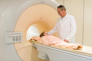
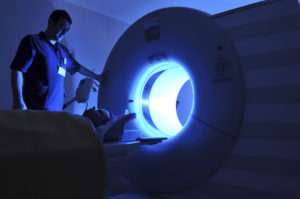
What can be expected when you have MR enterography?
As always, remember your basic MRI safety – no metal can enter the MR suite – this means all clothing with metal must be removed. MR enterography relies on adequate distention of the small bowel, usually using the same oral contrast agent containing iodine as for CT enterography. Images of the abdomen and pelvis will be obtained while IV contrast containing gadolinium is injected. The imaging time is longer for MRI than for CT, usually close to 30 minutes total for MR. Holding still is important as any motion will cause loss of detail.
What can we find with MR enterography?
This can show vascular lesions of the wall of the GI tract, masses and mucosal lesions as can be seen with inflammatory bowel disease. It also allows us to see detail in the bowel wall and in the adjacent soft tissues. Fistulas (abnormal communications from bowel loops), strictures and blockages, and abscesses can be seen in patients, often in those with inflammatory bowel disease. Problems with the blood vessels going to the bowel will be shown, such as narrowings or aneurysms.
And after the exam?
To flush the excess contrast from your system – drink lots of water! MR enterography is one more tool in the arsenal for imaging the small intestines able to produce beautiful images helping us keep you on the path to your best health.
Originally published 7/30/14 on diagnosticimagingcenters.com.