7 Things on a Typical Mammogram Report
 Happy Tuesday! It’s Day 8 of Breast Cancer Awareness Month! Today we’d like to talk about what might be included in a typical mammogram report.
Remember, reports can vary in length and lingo, as reporting styles are as individual as the doctors, but there is some standard content that helps them communicate your breast health to your referring clinician.
On a normal mammogram report you can expect some version of the following:
Title: This states what kind of imaging was done. Screening or diagnostic; bilateral (both breasts imaged) or unilateral (if just one side).
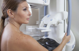
Technique: This will indicate how the mammogram was performed, with digital imaging (more accurate in some groups of women) or filmscreen (traditional); views taken may be listed with abbreviations which usually indicate the direction/angle of the view (standard views include MLO= diagonal and CC=top-down); implant displaced views will be listed (if applicable).
Indication: For normal reports this will be “screening” – as in, we are doing this to screen an asymptomatic woman for cancer. If a diagnostic study, any symptoms will be listed here. This section may discuss risk factors like family history.
Comparison: Will list the dates of available studies which were compared to the current. The more the better! Radiologists LOVE to have prior imaging to compare with current mammogram results. They are a great way to tell if there are any changes. (Did we say love….LOVE!)
Findings: Should include a statement of “parenchymal density” (that’s fancy-talk for what type of breast tissue you have); density can range on a scale from mostly fatty to mostly fibroglandular. This is where to look to find out if you have dense breast tissue. Here’s where your radiologist will mention things like symmetry or lack of changes in tissue (good things!). The report may list absence of suspicious things such as no suspicious calcifications, “architectural distortions” (fancy-talk for alteration of tissue) or masses. There may be a mention of benign findings, like lymph nodes in breast tissue, vascular calcifications, or stable (unchanged = good!) benign (noncancerous) nodules or masses. Small lymph nodes in the axilla (or armpit) are common and may perhaps get a mention here too.
Impression/Conclusion: Usually a statement that reads “negative” (<–that’s the good kind of negative, it means nothing was found) or benign. May give recommendations for follow-ups.
BI-RADS: A medical scale for identifying/explaining the findings and recommendations based on the imaging results. This helps your doctor understand the report and what to do next for best breast health.
We hope this short synopsis gives you a guide to navigating through the typical mammogram report – maybe now it will seem less like a document in a foreign language!
Image credit: in the paper by Chris Blakeley via Flickr Copyright Creative Commons Attribution-NonCommercial- NoDerivs 2.0 Generic (CC BY-NC-ND 2.0)
Originally published 10/8/13 on mammographykc.com.


