Breast MRIs and the Importance of Contrast
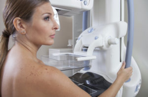
For breast imaging, mammography is the gold standard and the most familiar. Breast MRI is being used more and more, for lots of different reasons, including screening in some high risk patients. If we are doing a breast MRI to screen for cancer, to assess someone for the extent of breast cancer in her breast or as a problem-solving tool for abnormal findings on other tests, the study will be done with the help of intravenous (IV) contrast or dye. If we are doing a breast MRI to evaluate the structural integrity or intactness of breast implants or if the woman has poor kidney function, we will do the study without the IV contrast material.
So today, we’d like to take some time to talk about breast MRIs and why IV contrast may be a crucial part of the study.
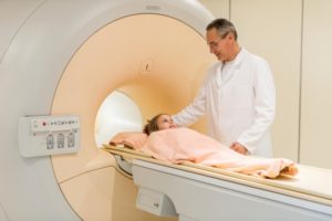
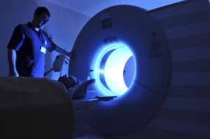
First, a quick run-down of an MRI. MRIs are large powerful magnets with images that are made by pulses of radiofrequency energy (really!!) that clank and clunk when the machine is running. Exam times usually run between 20-40 minutes. Over that time, hundreds of images of the body part being examined are generated. Motion can degrade the images, so getting as comfortable as possible initially and holding still are key.
If you come in for a breast MRI, you’ll be asked to change into a gown (and remove every scrap of metal from your body – important!). If you are being evaluated or screened for cancer, an IV will be started. You will lie facedown on the table. Your breasts will be adjusted to fit (comfortably) in a plastic device (called a coil, although not shaped like one!) specially made to create and make the images. Then it’s arms up over your head and the table slides into the magnet. Blankets are provided to keep you warm throughout the experience.
The images begin with sequences without contrast to look at the breast anatomy and show things like cysts and fluid collections. Implants will require special sequences to look at their internal structure. But there’s one more step when looking for cancer: contrast. Contrast agents for MRI are a liquid form of gadolinium, a heavy metal ion, that’s injected through an IV. Aside from the quick needlestick, it is a painless step, one where contrast diffuses into the bloodstream, illuminating the flow of blood in the images. When used for breast MRI, this step gives us powerful additional information on vessels and blood flow to normal and abnormal tissues in the breast. This goes beyond looking at just the anatomy, and picks up cancers that may not be visible by other means. This step creates even more images, numbering in the thousands, with radiologists often making use of computers to analyze some of the data.
Using IV contrast is a simple yet powerful tool in breast MRI. We hope this exploration of breast MRI helps in understanding the process and reasons behind the extra steps.
Originally published on 11/1/13 on mammographykc.com.


