Calcifications, in Detail
You’ve heard us broach the subject of microcalcifications in the past, but today we’d like to go further in depth on the topic. Calcifications in the breast are common- in fact the majority of mammograms will show some type of calcification, most often clearly benign and requiring no further evaluation. However, the smallest calcifications may be a sign of early breast cancer. Many invasive breast cancers will present with microcalcifications in addition to a mass. For these reasons, we’d like to explore the subject of microcalcifications further.
What are they?
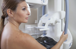
Microcalcifications are, as the name suggests, small deposits of calcium, a mineral, in the breast. Micro- refers to the size, with the small, millimeter size calcifications the ones we are most keen on finding. These are best seen on a mammogram – not as well evaluated on other breast imaging and one of the reasons mammography is supplemented by but not replaced by MRI. Once found, microcalcifications will be evaluated by the radiologist for signs that they are clearly benign (needing no further evaluation) or indeterminate or worrisome and needing further evaluation.
What causes them?
There are many benign, non-cancerous reasons for microcalcifications in the breast. Some of these include the following:
-
calcifications in the skin – these are often related to the sebaceous glands and if we are confident the calcifications are in skin on the mammogram can be sure they are benign;
-
fat necrosis – sometimes little areas of fat die – often from sort of trauma, either mechanical (being struck in the breast) or from surgery – and these may develop calcifications; the calcifications may be clearly benign and characteristic, but when first developing may be more problematic and require work-up;
-
fibrocystic changes – can also cause calcifications; these may be indeterminate;
-
vascular calcifications – these are another type of calcification which can fall in the microcalcification range – usually it is easy to see that they are related to the vessel wall, but occasionally may need further work-up to make sure that is where the calcifications are located;
As indicated above and why microcalcifications are searched for in every mammogram, microcalcifications can be a sign of breast cancer. If an isolated finding, this may be from ductal carcinoma in situ (DCIS). Many invasive cancers may also present with microcalcifications, often with a mass. These are the findings that make us more suspicious of microcalcifications:
-
irregular or branching shape
-
more than 5 calcifications grouped together
-
a distribution in the breast that is in a line or in a branching pattern (like it is in the duct)
-
new or increasing number of calcifications
-
microcalcifications with a mass
What is done next?
If the microcalcifications fall into one of the clearly benign, non-cancerous categories nothing beyond routine mammographic screening will be needed.
If clearly suspicious or indeterminate the following steps will often be taken:
-
compare to previous studies – if we can show they have been present and unchanged over multiple years, nothing further may need to be done;
-
magnification spot views – you may be recalled from a screening mammogram for further work-up which for calcifications will often include views focused on the area in question done with magnification technique; this lets us see the calcifications better, will show us the extent of the calcifications more accurately, and often will allow us to put them into either clearly benign or clearly suspicious categories; some will remain indeterminate
-
if suspicious in character, a biopsy will be recommended;
-
if indeterminate and not an unchanged finding, biopsy will likely be recommended;
-
if the history and appearance suggest these may be benign, as from fat necrosis, we may recommend a short term follow-up mammogram, most often in 6 months;
-
if there is an associated mass, we will likely do an ultrasound;
Remember, even when biopsy is recommended, there is still a strong likelihood that the calcifications will be benign – we don’t yet have a way to determine which of those indeterminate groups we can ignore. If we look at all patients with biopsies for microcalcifications, the majority, 80-85% will have benign results on their biopsy.
We know that any finding not in the negative or benign category on mammography can lead to confusion and stress. Microcalcifications can definitely fall in that category, and we hope this more in-depth exploration will answer questions and reduce stress.
Originally published 12/9/13 on mammographykc.com.


