CT Ent-enter-entero-enterography! Whew!
 When investigating issues of the abdomen and intestines, your doctor may order a small bowel series or enteroclysis. However, CT technology is also an excellent noninvasive way of looking at internal organs. It also allows us to better see the tissues adjacent to the small bowel. If a dedicated view of the bowel and adjacent soft tissues is needed there is a special procedure called CT enterography which may be used.
Why would you need CT enterography?
There are many potential reasons why a patient may come in for CT enterography, with the goal of precisely defining anatomy and potential causes of symptoms. Symptoms can include:
- Abdominal pain (especially in the left or right lower quadrant)
- Blood in stool
- Possible bowel obstruction, usually partial blockages of the small bowel which can be difficult to diagnose and difficult to find the cause of
- Inflammatory bowel disease – with CT enterography we can see not just the disease of the bowel, but any complications in adjacent tissues which are frequent
- Hernias
- Masses
- Narrowings or strictures
- Infections
- Masses in the mesentery or adjacent organs that can affect the GI tract
How does a patient prepare for CT enterography?
Patient prep for this type of imaging is fairly basic: fasting (no food or drink) for 4 hours before the CT may be requested. At times the study may be done without preliminary fasting.
What can be expected when you have CT enterography?
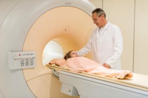
Typically, CT enterography uses fairly large volumes of oral contrast agents which are iodine-based and iodine-based IV contrast. The oral contrast material helps distend the bowel, so that the wall is well seen. The blood vessels and any inflammatory changes are best seen with the IV contrast highlighting the vessels. If you have allergies, let us know so we can plan ahead. CT uses radiation, so this exam in general is not done in those that are pregnant or who may be pregnant.
What might we see with CT enterography?
- Inflammatory bowel disease – bowel wall thickening, abnormal enhancement of the wall of the bowel, strictures, fistulas (abnormal communication between bowel loops), adjacent abscesses all may be signs of inflammatory bowel disease and imaging with CT enterography can help assess its degree of activity
- Masses – benign polyps and cancers
- Vascular problems to the small or large bowel, including narrowings of vessels or aneurysms (small saccular outpouchings)
- Blockages – we can not only see the site of obstruction but many times can find the cause of the blockage
- Infections
CT enterography is a fusion of the best of CT technology, allowing us to see the soft tissues in the abdomen, and enterography which distends the small bowel, allowing us to best see the wall. With this technique, small bowel diseases are beautifully demonstrated.
Originally published 7/23/14 on diagnosticimagingcenterskc.com.
