Head Aches and Head Issues #4: Head MRI – What To Expect
 If you are experiencing headaches and your evaluation by your doctor suggests the need for imaging, you may be sent for either an MRI or CT scan. CTs are quick and valuable in the evaluation of patients presenting with headaches after trauma. MRI is an alternative means of imaging the brain and adjacent tissues. CT vs. MRI The main differences in the two technologies are as follows. The decision as to which test is needed is based on your history and findings, as well as the following:
CT:
- uses ionizing radiation (avoided in pregnancy unless there are significant findings or significant trauma) in conjunction with computers to generate images
- takes 10 minutes or less
- may or may not use IV iodinated contrast material
- great in looking for blood in and around the brain, which can be traumatic or non-traumatic in origin
- uses a short bored tube
MRI:
- uses magnets and radiofrequency waves in conjunction with computers to generate images – no radiation
- can be used in pregnancy after the first trimester and without IV contrast material
- may or may not use IV gadolinium contrast material
- takes 30 minutes or more
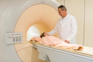
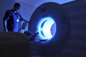
- uses a long bore tube (can seem confining although there are ways of treating this sensation!)
- shows anatomy in greater detail than CT
- some pathologies such as multiple sclerosis are best visualized on MRI
- must hold still for longer time periods – may be difficult for younger children
What To Expect
Before an MRI of the head, no special preparation is necessary. However, metal is a big issue (seriously, the machine is one giant magnet and any metal on your body can become a hazardous missile with potential for harm to you, the technologist or the machine). So – extreme care is used to ensure that you have no metal on your body. Also, metals in things like artificial joints and pacemakers can create problems so full disclosure is needed. The procedure takes approximately 30 minutes with only the head moving through the machine. Holding still during the imaging is key to getting good pictures. Images are taken without contrast to begin with and then if needed (and patient is not pregnant) additional series may be run after an IV injection of a contrast material containing the heavy metal gadolinium. This should be used in caution in certain patients with kidney problems, so we always obtain a full history prior to giving this, and may check your kidney function before giving it. The injection may cause a cooling sensation.
What Happens Next
After your exam is completed, the images are studied by your radiologist for interpretation and reporting. The results are then shared with your referring doctor to integrate the new information gained from your head MRI with clinical symptoms for a specific diagnosis. After the test, we recommend drinking extra fluids to help flush the contrast from your system if it was used.
On Your Way!
Headaches can be a vexing issue, and getting you on the road to being headache-free is the goal of the medical team, including the radiologist carefully analyzing those images. As ever, we hope to help to get you on the road to your best possible health.
Originally published 8/22/14 on diagnosticimagingcenterskc.com.
