Imaging of the Lumbar Spine
 To continue our conversation about back pain, today we’re going to focus on the different imaging tests that may be requested to evaluate someone with back pain, specifically lower back pain. Imaging for lower back pain will likely focus on the lumbar spine. It’s the lower back region, usually the lower 5 vertebral levels – the series of vertebrae highlighted in the picture here.
There are several modalities we use to examine the lumbar spine, including (but not limited to) x-ray, MRI and CT. Conventional films or X-rays are often the starting point when evaluating someone with back pain. This allows an overall assessment of alignment and can look for signs of a more serious issue requiring additional imaging, like a fracture or an area of abnormal appearing bone. If the back pain follows recent trauma, is seen in someone with known osteoporosis or is in someone over the age of 70, conventional films may be the imaging study of choice and may be the only imaging needed.
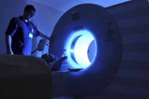
MRI (magnetic resonance imaging) of the lumbar spine is the most common imaging test for back pain because of its ability to see details of the soft tissues, including the nerves as well as the bones.
As an added benefit, no radiation is involved. Remember with MRI the images are made with a large magnet and radiofrequency pulses (those are the loud sounds you hear). With MRI we can examine all of the following: the structure of the discs (the cartilage cushions between the vertebrae) and look for how the disc relates to adjacent nerve roots; the bones that make up the spine including the vertebral bodies; the conus (the lower end of the spinal cord); and the soft tissues surrounding the area. Imaging takes about 30 minutes, and remember – you can’t have an MRI if you have a pacemaker (most types anyway!) or some implanted devices like stimulators. Lastly for the safety of all you cannot bring metal of any kind into the room!
CT (computed tomography) is an additional means of looking at the spine. Images with CT are made with x-rays that are created in a ring around the body allowing the body to be imaged in thin slices. CT allows an excellent look at the bones making up the spine. It may be the study of choice for evaluation of acute trauma or fracture. Changes in the discs can be seen, but the nerve roots are not visualized in a routine CT. There are times when a CT of the spine is performed after a myelogram – this is a procedure where contrast material is put in the fluid- containing space surrounding the spinal cord and nerve roots. This then allows CT to better evaluate the nerve roots and soft tissues of the spinal canal. A CT scan may be used to help with planning before back surgery. A CT lumbar spine takes around 10 minutes. It should not be used in those who are pregnant unless it is an emergency (as with severe trauma).
All of these are straightforward procedures that can give an excellent anatomic evaluation. Remember imaging of the spine will often reveal age-related changes in the discs and bones, not all of which will be associated with symptoms. Imaging is not always needed for back pain – see your doctor to determine if imaging can help get you on the path to your best possible health!
Image credit: Lumbar vertebrae anterior by Anatomography via Wikimedia Commons Copyright Creative CommonsAttribution-Share Alike 2.1 Japan
Originally published 5/8/14 on diagnosticimagingcenterskc.com.


